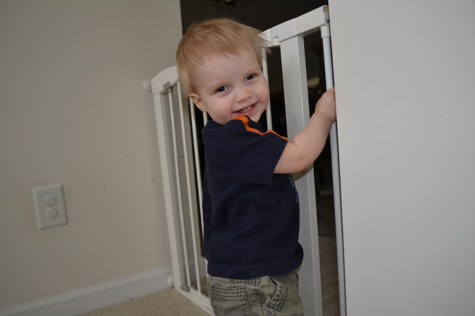
The Exam
Alex had his
first exam under anesthesia (EUA) today.
He did really well. In the triage
area, we read a lot of books and tried to keep him on our laps as much as
possible. The walls were painted with a
farm scene and Alex petted the cows and pretended the bees were stinging him. He was given two sets of eye drops to dilate his eyes.
Things we didn’t
expect:
How long it
would take. Our appointment time was
8:45 am (we got there at 8:15am) and Alex was discharged at 1pm. Parents – bring snacks for yourself!
How many
diapers we would need. Between the last
apple juice 2 hours before his appointment time, all of the IV fluids, and lots
more apple juice after the exam – he needed lots of diaper changes.
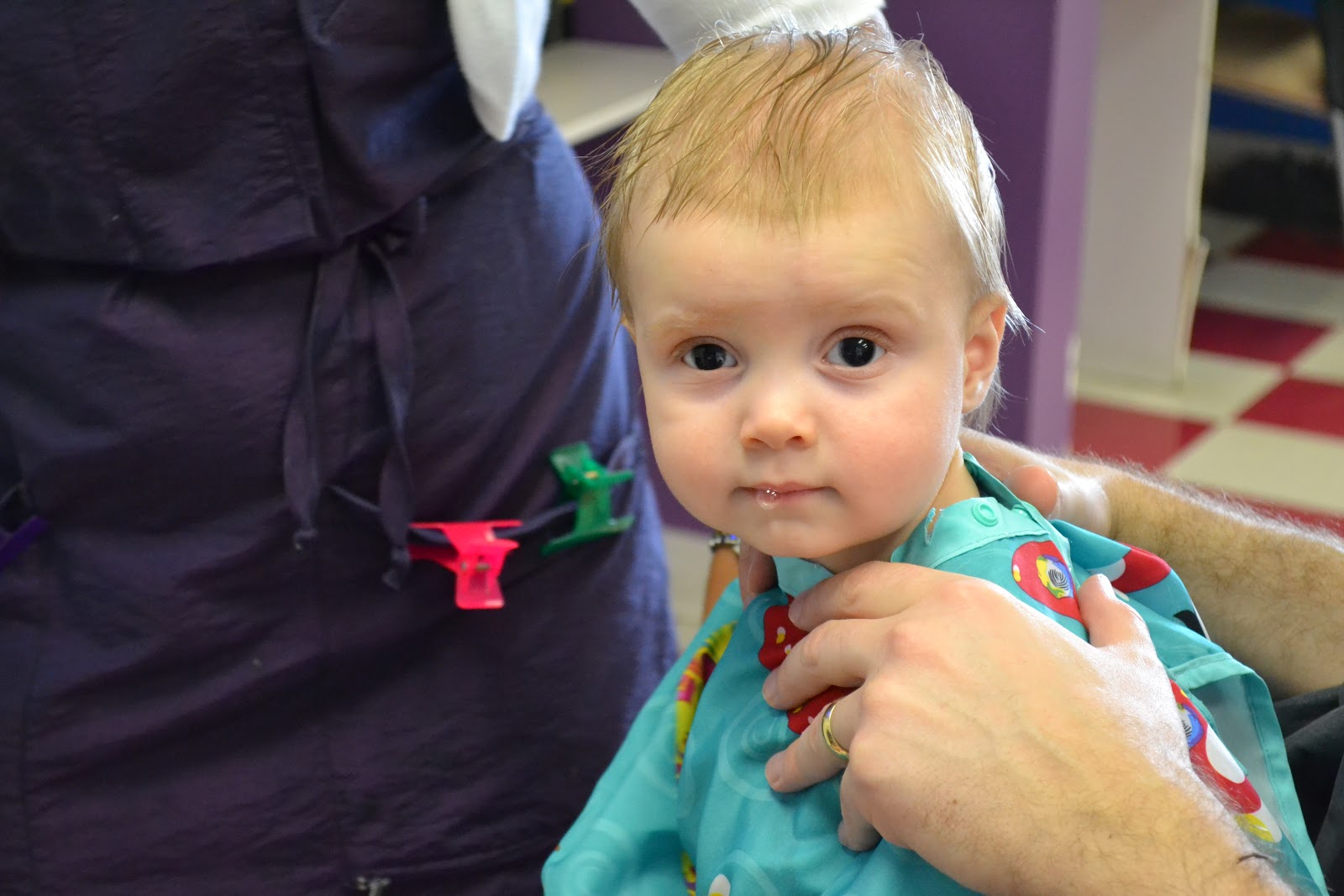
What we
would need in the recovery room – diapers, sippy cup, clothes to redress. The nurse had to find Rob to bring those
things to me.
His eyes to be sensitive afterward. We took Alex's sunglasses off when we got home, but he grabbed them and put them back on. He wore his sunglasses until bedtime.


Since he is
so young, the anesthesiologist recommended that Alex take an oral sedative so
that it would be easier to apply the gas.
It doesn’t taste very good, but he didn’t spit it out. He just made faces and tried to touch his
mouth. We kept him busy by having him
point at things around the room. I have
a feeling this trick won’t work at his next EUA. After Alex had the oral sedative, he started
to get a bit drowsy. His head flopped
and his speech was a little slower. Rob
gowned up and we waited until it was time to take Alex back. Rob went with him when they applied the gas
and I waited in the triage area.

The EUA took
about 45 minutes and we waited in the
pediatric waiting room. Alex’s doctor
met us in the waiting room and let us know how the exam went. I went back to the recovery room and waited
for Alex to wake up. He wasn’t waking up
on his own so the nurse had to wake him up.
He was groggy and cranky. We went
back to the triage area and Alex had some apple juice and graham crackers. Rob joined us and dressed Alex while he ate
and drank his snack while sitting on my lap.
Results
I am
summarizing below from the notes I scribbled on the back my Duke Eye Center
word search. We need to do some research
to get a better understanding and digest everything.
The Good:
The Not So
Good (indicators Alex is at risk for Glaucoma):
So what’s
next?
“In the past decade, many surgical advances were made in the
field of ophthalmology and some of these surgical treatments were attempted on
individuals with aniridia. As
we continue to study aniridia, we are learning is that the aniridic eye is very
delicate and any intraocular surgery is risky because of the impact on the
other structures of the eye. Many times, a treatment to address
one issue can result in another issue (for example, corneal surgery that fixes
the cornea but results in glaucoma. Or, glaucoma surgery that results in damage
to the limbus region or a cataract.) Also, a condition called
aniridia fibrosis where fibrotic tissue begins to invade the interior of the
eye.” – Vision for Tomorrow website
We need some
time to think about all of this and do some research.
Right now we
are so happy to have our little boy back home.
The nurse said he would be tired and want to sleep. Not so!
Alex told Rob he wanted to, “Run!” around the playroom.
“Glaucoma. Glaucoma is a group of eye diseases that gradually steal sight by harming the optic nerve. In the early stages of the disease, there may be no symptoms. In most cases, glaucoma is caused by an increased pressure within the eye. This elevated pressure is caused by a backup of fluid in the eye. Over time, it causes damage to the optic nerve.
In order to best understand what happens in glaucoma, think of the eye as a sink, in which the faucet is always running and the drain is always open. The aqueous humor (the fluid) is constantly circulating through the anterior chamber. This fluid is produced by a tiny gland, called the ciliary body, situated behind the iris. It flows between the iris and the lens and, after nourishing the cornea and lens, flows out through a very tiny spongy tissue, only one-fiftieth of an inch wide, called the trabecular meshwork, which serves as the drain of the eye. The trabecular meshwork is situated in the angle where the iris and cornea meet. When this drain becomes clogged, aqueous cannot leave the eye as fast as it is produced, causing the fluid to back up. However, because the eye is a closed compartment, the “sink” does not overflow; instead, the backed up fluid causes increased pressure to build up within the eye.
To understand how this increased pressure affects the eye, think of your eye as a balloon. When too much air is blown into the balloon, the pressure builds, causing the balloon to pop. Because the eye is too strong to pop, it gives at the weakest point, which is the site in the sclera where the optic nerve leaves the eye. As we mentioned above, the optic nerve is the part of the eye which carries visual information to the brain. It is made up of over one million very thin nerve cells. When the pressure in the eye builds, the nerve cells become compressed, causing them to become damaged and, eventually, die. The death of these cells results in permanent visual loss.”


